[ad_1]
Introduction
CT and MRI are important preoperative imaging investigations for bone and tumor info for surgical planning in orthopedic oncology. As attaining unfavorable margins in tumor resections decide the oncological outcomes1, spatial understanding of the patho-anatomy is essential for the success of tumor resections in numerous sufferers with distinctive anatomies and tumor extents.
Conventionally, tumor surgeons view two-dimensional (2D) medical photographs in a pc station. Image processing software program could create three-dimensional (3D) fashions from 2D imaging datasets. Visualizing digital 3D fashions on the 2D flat display of the pc station lacks depth notion and parallax in comparison with bodily 3D fashions. When surgeons clinically study bone tumor sufferers earlier than the surgical procedures, they need to mentally course of and accurately overlay the sufferers’ digital 2D medical photographs and 3D bone-tumor fashions onto their anatomy. Also, surgeons should mentally retain the spatial orientation of the matched digital 3D fashions when viewing sufferers from totally different angles. Appropriate surgical exposures and the osteotomies alongside desired planes are then determined. The visual-spatial capability (VSA) to mentally visualize and manipulate objects in 3D house is extremely demanding,2 particularly when the quantity of concurrently processed info is substantial for big tumors, areas with complicated anatomies, and bone geometries.
Computer navigation and 3D-printed resection guides facilitate exact osteotomies3,4 however primarily profit orthopaedic procedures after surgical publicity. Computer navigation permits monitoring of the diseased bones on the subject of the preoperative medical photographs after inserting a navigation tracker into the affected person’s bone. Also, surgeons function by viewing the 2D photographs within the navigation show and are distracted from the operative fields.5 Although bodily 3D-printed fashions could present a viable various for visualizing the affected person’s patho-anatomy with tactile suggestions,3 psychological translation of the spatial relationship of bodily 3D fashions to the precise sufferers’ anatomy continues to be required. Both applied sciences could not overcome the surgeons’ cognitive load within the spatial understanding of bone tumors in precise sufferers earlier than the surgical procedure begins.
Mixed Reality (MR) is an immersive know-how with spatial computing in merging actual and digital worlds, and customers can work together with digital objects in actual time.6 The digital digital objects in MR are 3D holograms that retain the depth and parallax notion of the unique bodily objects. Through Head-Mounted Displays (HMDs), surgeons stereoscopically view the 3D holograms from 360° of their bodily atmosphere. Patient-specific 3D holograms are generated from 2D medical photographs. By superimposing the 3D holograms on the affected person, surgeons can intuitively perceive the spatial relationship of bone tumors to the affected person’s anatomy. Clinical stories of MR utility are restricted, with only some on backbone pedicle screw placement7 and shoulder arthroplasty.8 The information in orthopaedic oncology is missing. In this examine, we evaluated 1) surgeons’ spatial consciousness of tumor areas and orientations by viewing patient-specific 3D holograms; 2) the cognitive load of surgeons when utilizing MR know-how for preoperative medical evaluation of bone tumor sufferers.
Methods
The examine was carried out beneath the moral requirements of the Joint Chinese University of Hong Kong-New Territories East Cluster Clinical Research Ethics Committee (the Joint CUHK-NTEC CREC) (CRE Ref. No. 2022.543) of the authors’ hospital. Informed verbal consent was obtained from the sufferers collaborating within the examine. The verbal consent as a substitute of written consent was authorized by the Joint CUHK-NTEC CREC. The examine complied with the Declaration of Helsinki. Between July 2021 and December 2022, we retrospectively reviewed 9 sufferers with major bone tumors having surgical procedure. There have been six major bone sarcomas, two benign bone tumors, and one revision pelvic prosthesis (Table 1). The tumor areas have been pelvis (three), tibia (two), proximal femur (one), scapula (one), proximal humerus (one), and calcaneus (one). All sufferers (besides Patient 1) underwent bone resections with 5 sufferers beneath the help of 3D-printed resection guides. Three sufferers had 3D-printed patient-specific implants for bone reconstruction, one with iliac crest bone graft, two with cementation, and no reconstruction in two sufferers. Surgeons used MR know-how for preoperative medical evaluation. MR know-how was thought mandatory for preoperative medical analysis of bone tumor sufferers due to anticipated difficulties in spatial understanding of complicated anatomical areas such because the pelvis, intraosseous tumor involvement, websites of deliberate osteotomies, or the position of patient-specific guides and implants (Figure 1A–H).
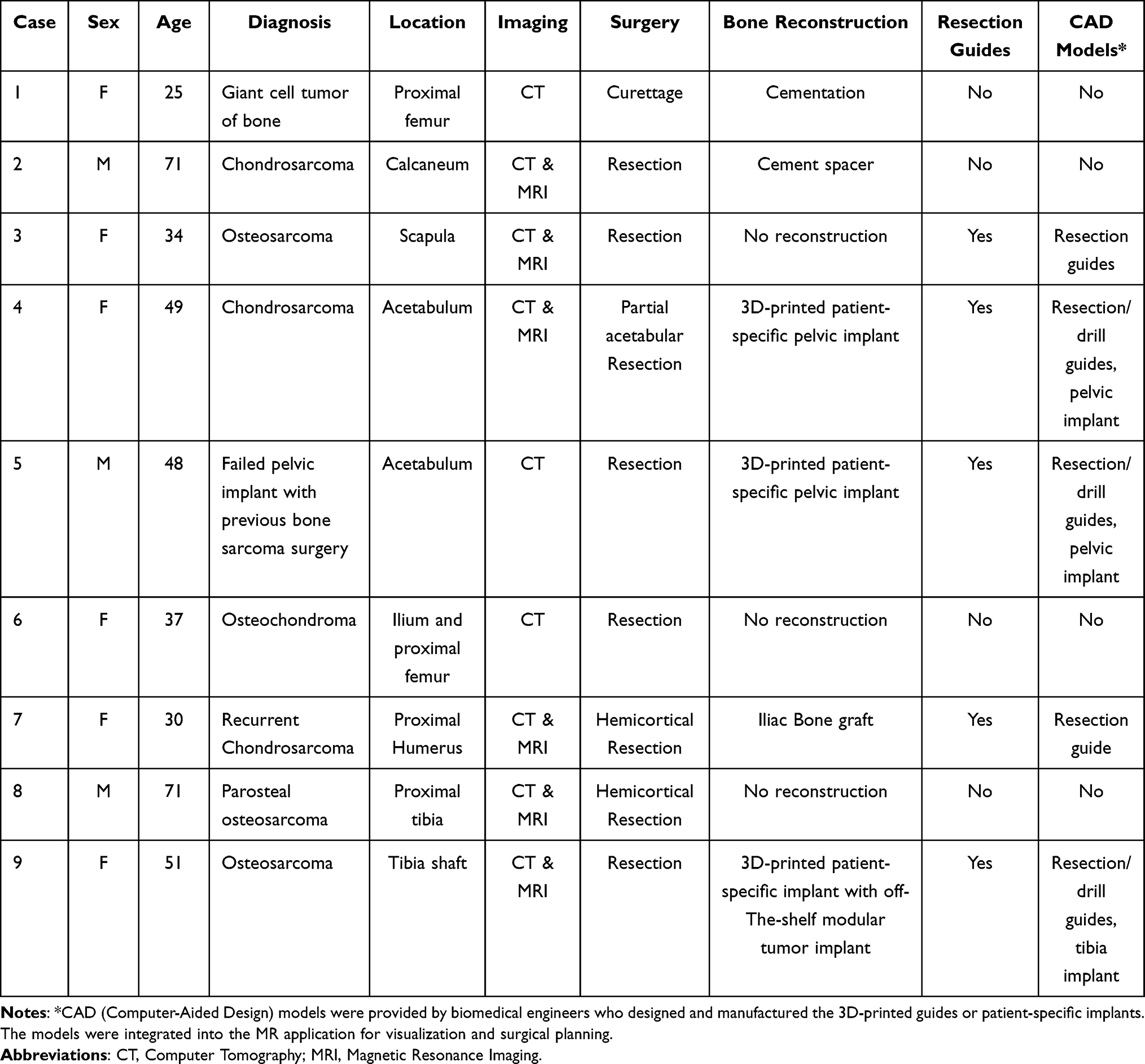 |
Table 1 Demographic Information of the Patients Using Mixed Reality Technology within the Study |
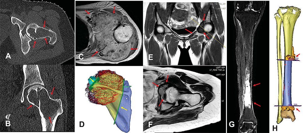 |
Figure 1 The axial (A) and coronal (B) views of CT photographs in Patient 1 with large cell tumor involving femoral head and neck (pink arrows). The axial view (C) of T1-weighted MRI with distinction and 3D surgical planning (D) with patient-specific chopping guides (yellow arrows) in Patient 3 with left scapular osteosarcoma (pink arrows). The coronal view (E) of T1-weighted MRI and axial view (F) of Proton Density sequence MRI in Patient 4 with left acetabular low-grade chondrosarcoma (pink arrows). The coronal view (G) of T2 weighted MRI in Patient 9 with tibia osteosarcoma (pink arrows) and 3D surgical planning (H) with patient-specific chopping guides (pink arrows). |
Preparation of MR Holographic Application
The medical workflow of blended Reality in orthopedic oncology has been described9 (Figure 2). The MR software program platform was developed (Syngular Technology Limited, Hong Kong SAR, China), and MR holographic utility was ready for every case. CT ± MRI photographs have been acquired as each are important preoperative imaging investigations for the workup of musculoskeletal tumors. The 2D medical photographs in DICOM format have been obtained for additional processing. 3D bone fashions have been generated from CT photographs as bones have a excessive distinction sign. MRI photographs higher present delicate tissue buildings, and medullary and extraosseous tumor volumes have been mapped. 3D bone-tumor fashions have been then created with their geometries represented in polygons. Polygon-based Computer-Aided Design (CAD) fashions have been thus produced from 2D medical photographs. Computer Graphics software program generates cinematic-rendered (CR) fashions with edited colour, materials, texture, and the suitable illumination to simulate the pure manner mild would replicate off of bodily objects in the actual world. The processing offers CR fashions a extra photorealistic depiction of the 3D fashions (Figure 3A–E). A 3D Engine (Unity Technologies, Unity Software Inc., San Francisco, US), a system generally used for digital laptop simulations, was then used to develop the User Interface (UI) for the holographic contents, together with 2D medical photographs, digital medical info, and 3D fashions. The holographic utility calculates the visible look of the scene, and the interactive 3D holograms have been then created. Its content material was exported right into a digital package deal and loaded into the MR-HMD (Hololens 2, Microsoft Corporation, Redmond, WA, US).
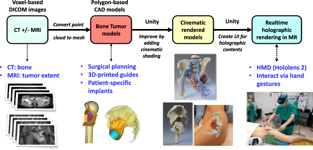 |
Figure 2 Shows the workflow of producing the blended actuality software program utility used for surgical planning of bone tumors. Abbreviations: DICOM, Digital Imaging and Communications in Medicine; CT, Computer Tomography; MRI, Magnetic Resonance Imaging; CAD, Computer-Aided Design; UI, User Interface; MR, Mixed Reality; HMD, Head-Mounted Display. |
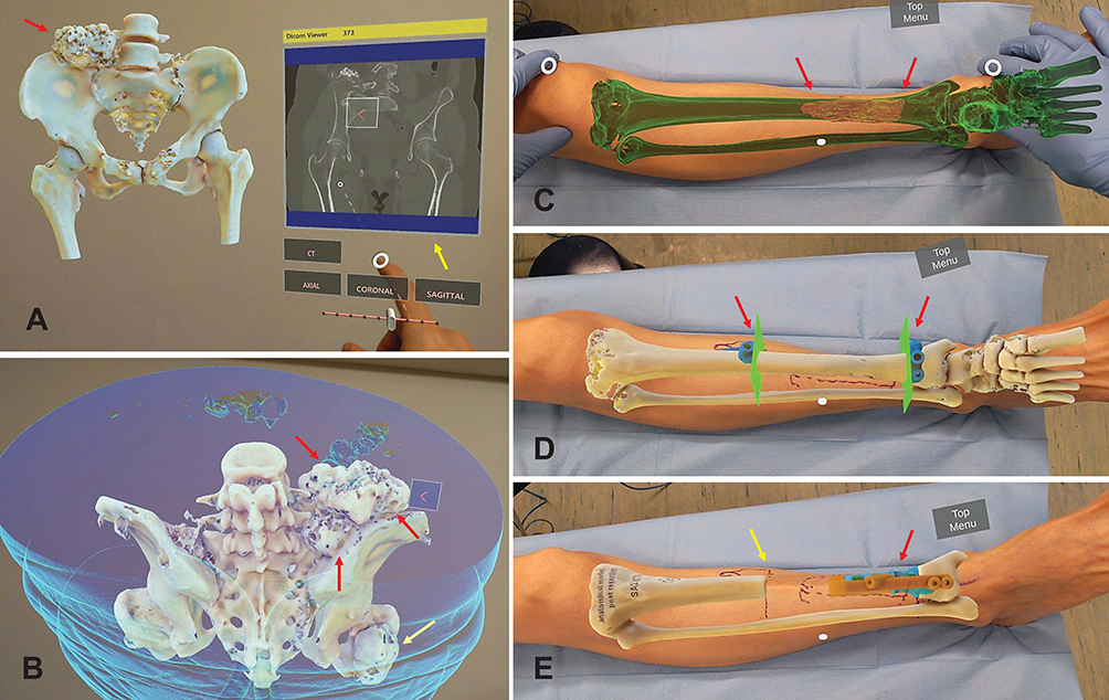 |
Figure 3 (A) reveals cinematic-rendered 3D holograms of the pelvis and proximal femur in Patient 6 with the osteochondroma of the precise ilium (pink arrow). Using hand gestures, the surgeon interacts with the 3D holograms and the 2D picture of the CT pelvis (yellow arrow). The entire picture datasets in axial, reformatted coronal, and sagittal views will be considered and scrolled by in real-time. (B) reveals the superior and posterior views of the pelvic and Hip 3D holograms in Patient 6 with osteochondroma of the precise ilium (pink arrows) and proximal femur (yellow arrow). (C) After matching the cinematic-rendered 3D hologram of the tibia osteosarcoma to the leg of Patient 9, the surgeon examines by visualizing the osteosarcoma (pink arrows) with an improved spatial understanding of tumor boundaries. (D) The resection planes and patient-specific chopping guides (pink arrows) may also be built-in into the holograms that facilitate the resection planning and positioning of the chopping guides. (E) The bone reconstruction with a patient-specific implant and its respective drill information for screw fixation (pink arrow) will be visualized. The resected tibia bone hologram allows the surgeon to mark the osteotomy web site (yellow arrow) exactly earlier than the pores and skin incision. The blended actuality hologram visualization on patents enhances surgeons’ capability for complicated surgical planning. |
Surgical Planning and Clinical Examination
The surgeon (KCW) carried out the surgical planning by clinically accessing every bone tumor affected person utilizing standard 2D and MR 3D hologram strategies. In the standard 2D technique, the surgeon reviewed the 2D picture information, planning info of 3D-printed guides and patient-specific implants (if any), then mentally overlaid the digital 3D fashions onto the affected person’s physique (Figures 4A, B, D–F; 5A–C, E and F). In the MR 3D hologram technique, the surgeon visualized the 3D holograms instantly on the sufferers by way of HMD. The HMD incorporates cameras that monitor the palms of the surgeons. Hand gestures have been used for real-time interplay with the 3D holograms. Surgeons manually matched the holograms onto the affected person’s anatomy. The registration was verified as appropriate by palpating the close by bony landmarks in pelvis (anterior superior iliac backbone, posterior superior iliac backbone or pubic tubercle); tibia (medial and lateral malleoli or tibial tuberosity); humerus/scapula (higher tuberosity, clavicle, acromion or scapular backbone); calcaneus (calcaneal tuberosity). Then, surgeons “saw-through” the affected person’s physique with superimposed 2D medical photographs and 3D fashions (Figure 4C and G, Supplementary Videos 1 and 2). The surgical incision, publicity, websites of osteotomies, 3D-printed guides, and patient-specific implants have been then decided whereas clinically accessing every affected person with each strategies (Figure 5D, G and H, Supplementary Videos 3 and 4).
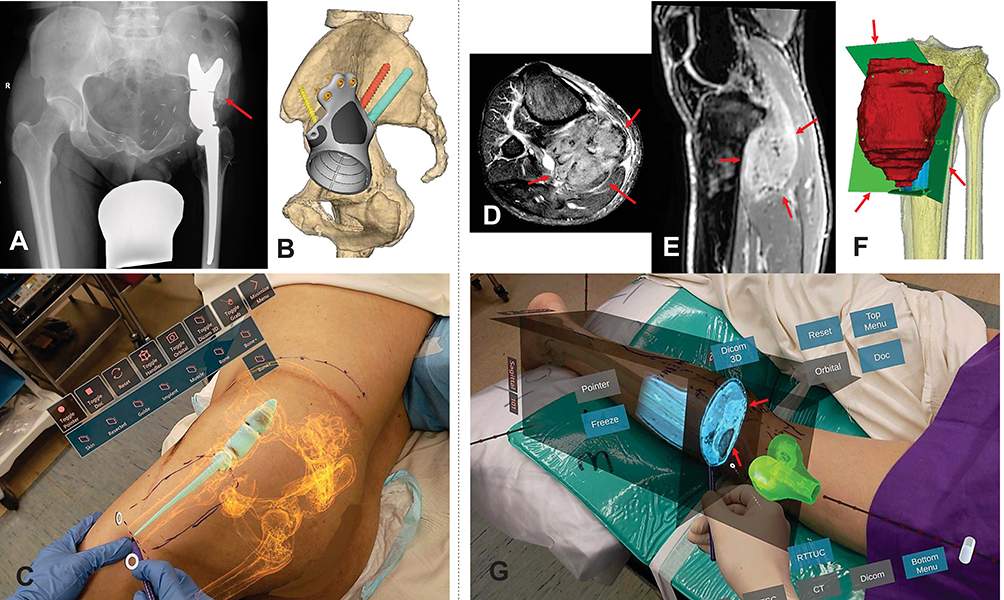 |
Figure 4 Patient 5 had left pelvic low-grade bone sarcoma with resection and saddle prosthesis reconstruction in 2005. He introduced with growing again ache and ipsilateral knee ache resulting from restricted hip movement. (A) reveals the plain radiograph of the pelvis with a saddle prosthesis with pseudo articulation at the next hip heart. Bone overgrows across the saddle element (pink arrow). (B) reveals the surgical planning of bone resection on the ilium and reconstruction of a patient-specific implant with customary hip arthroplasty on the close to regular hip heart. (C) By visualizing the 3D hologram on Patient 5 with distorted pelvic anatomy like X-ray imaginative and prescient, the surgeon “sees through” the pores and skin and determines the placement of the outdated implant, the surgical strategy, and the pores and skin incision web site in planning the revision surgical procedure (Supplementary Video 1). Patient 8 had proper posterior proximal tibia parosteal osteosarcoma with a big extraosseous tumor element (pink arrows) proven on the axial (D) and sagittal (E) views of MRI photographs. (F) reveals the surgical planning of the hemicortical tumor resection within the 3D bone tumor mannequin with deliberate resection planes (pink arrows). (G) As MRI photographs higher delineate the tumor extent (pink arrows) and delicate tissue buildings, the blended actuality characteristic of superimposing MRI picture dataset additional will increase the surgeon’s capability to grasp the spatial relationship of bone tumors for surgical planning earlier than pores and skin incision (Supplementary Video 2). |
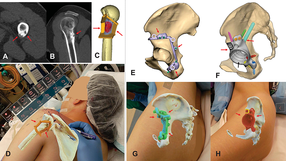 |
Figure 5 The axial (A) and sagittal (B) views of CT photographs present recurrent low-grade chondrosarcoma involving the posterior proximal humerus (pink arrows) in Patient 7. (C) reveals the 3D surgical planning of the hemicortical tumor resection assisted by a patient-specific chopping information (pink arrows). (D) Viewing the overlaid 3D holograms (bone and chopping information) on Patient 7 permits straightforward marking of pores and skin incisions and offers higher spatial consciousness of the chopping information placement (pink arrows) (Supplementary Video 3). (E) reveals the 3D surgical planning of partial acetabular resection assisted by a patient-specific chopping information (pink arrows) in Patient 4 with low-grade chondrosarcoma. (F) reveals the planning of reconstruction of the acetabular defect with a patient-specific implant (pink arrows) in the identical Patient 4. A twin mobility cup was cemented into the acetabular cup (black arrow) of the pelvic implant. 3D holograms of patient-specific chopping guides (G) (pink arrows) and implants (H) (pink arrows) may very well be visualized for surgical planning earlier than pores and skin incisions (Supplementary Video 4). |
No quantitative instrument was obtainable to evaluate the customers’ expertise in spatial consciousness of bone tumors throughout preoperative evaluation. Therefore, for every technique, the surgeon accomplished a qualitative survey 1) a Likert-Scale (LS) questionnaire to evaluate his opinions on the spatial consciousness of the bone tumors and the effectiveness of surgical planning and a couple of) The National Aeronautics and Space Administration-Task Load Index (NASA-TLX) to commute the surgeons’ subjective cognitive workload.10,11 The LS questionnaire makes use of five-point score scales to evaluate the surgeons on 5 particular domains: spatial visualization, spatial orientation, depth notion, identification of the placement of the pathology, ease of figuring out surgical incision, and effectiveness as a preoperative evaluation instrument. Spatial visualization is the power to have a look at a 2D determine and visualize what it could appear to be expanded into 3D, then mentally rotate and manipulate objects with out the bodily objects in entrance of you.12 Spatial orientation is the power to determine objects’ place or course in house mentally.13 The LS questionnaire was modified from the earlier examine by Lu et al.10 It had a five-point scale the place “1” pertains to a unfavorable expertise, and “5” is the utmost diploma of satisfaction. NASA-TLX is a measuring instrument to find out an general cognitive workload score after the surgeon performs the duty. The surgeon rated his rating on an interval scale between 0 and 100 on six subscales.14,15 The increased rating signifies a extra important workload of the respective subscales. The six parameters are 1) Mental demand – how a lot pondering, deciding, or calculating was required?; 2) Physical demand – The quantity and depth of bodily exercise required; 3) Temporal demand – the period of time stress concerned; 4) Effort – how arduous did you’re employed to take care of your degree of efficiency?), 5) Performance – the extent of success in finishing the duty, and 6) Frustration degree – How insecure, discouraged, or safe or content material you felt throughout the activity.
All resected bone tumor specimens have been examined histologically, and 6 sufferers with bone sarcoma have been additionally assessed for surgical margins.
Statistical Analysis
The outcomes of the LS questionnaire and NASA-TLX rating utilizing standard 2D and MR 3D hologram strategies for preoperative surgical planning in bone tumor sufferers have been recorded and in contrast. The outcomes have been summarized within the median and interquartile vary (IQR). The scores of two strategies (2D vs MR 3D hologram) in every area have been visually in contrast utilizing boxplots. Data analyses have been carried out utilizing R 4.2.1 (R Foundation for Statistical Computing, Vienna, Austria).
Results
The qualitative survey based mostly on the LS questionnaire revealed that the MR 3D hologram group appeared to have superior leads to all domains of spatial consciousness of tumors and was thought-about simpler as a preoperative planning instrument than the standard 2D Group (Table 2, Figure 6A). For NASA-TLX scores, the general cognitive workload was decrease in MR 3D hologram group than within the 2D Group for preoperative medical evaluation (2D group median rating 75, IQR 21.25–68.75 versus MR 3D group median 17.5, IQR 20–42.5). It is obvious that “Mental Demand”, “Effort”, and “Frustration” have been the dominant supply of cognitive workload within the standard 2D Group, whereas “Physical Demand” was within the MR 3D hologram group (Table 2, Figure 6B). When utilizing MR know-how with HMDs, the surgeon reported no discomfort, like movement illness, headache, fatigue, or eye pressure.
 |
Table 2 Results of Likert Scale Questionnaire Scores and NASA Task Load Index Scores |
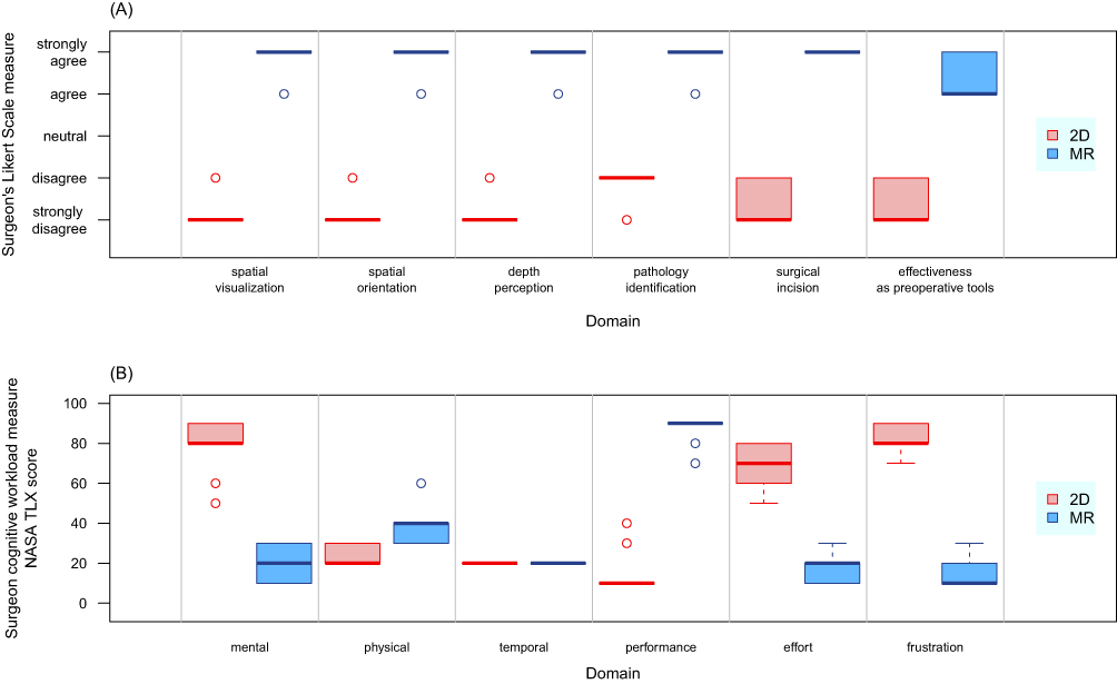 |
Figure 6 Shows the qualitative outcomes of a Likert-Scale (LS) questionnaire (A) on the spatial consciousness of bone tumors and the effectiveness of surgical planning and the National Aeronautics and Space Administration-Task Load Index (NASA-TLX) rating (B) on the surgeon’s subjective cognitive workload utilizing blended actuality know-how. Abbreviations: 2D, Conventional technique of viewing two-dimensional medical photographs and planning info; MR, surgical planning by viewing three-dimensional holograms that have been superimposed on the sufferers. |
Histological examination of the resected specimens confirmed that two of the six bone sarcoma sufferers had optimistic microscopic margins within the delicate tissue. All bone resection margins have been unfavorable.
Discussion
Surgical planning is difficult in complicated bone tumor surgical procedure. Surgeons should mentally combine preoperative 2D photographs and superimpose digital 3D fashions onto the affected person’s anatomy to find out the surgical strategy and osteotomies across the bone tumors. Although laptop navigation and 3D-printed guides allow exact osteotomies, the applied sciences are solely possible after surgical publicity. Our preliminary outcome advised that when put next with the standard 2D technique, MR 3D holograms could enhance 3D visualization and spatial consciousness of bone tumors within the affected person’s anatomy with out a rise in cognitive workload throughout the preoperative surgical evaluation. The new know-how could facilitate surgical planning earlier than pores and skin incisions in orthopaedic oncology surgical procedure.
The examine has a number of limitations. 1) It solely included a small pattern measurement (9 instances) that didn’t permit for significant statistical evaluation. A bigger variety of sufferers is required to conclude the statistical significance of potential superior leads to blended actuality know-how. 2) It is the expertise of a single orthopaedic oncology surgeon utilizing blended actuality know-how. Multiple surgeons are required for comparability to cut back assessors’ bias in future research. 3) Improved spatial consciousness of the tumor areas or orientations in preoperative medical evaluation utilizing MR could not translate into higher medical outcomes ruled by different components like tumor grades, response to chemotherapy, availability of assistive instruments to duplicate surgical plans intraoperatively, or resection margins. 4) Mixed actuality know-how continues to be creating within the orthopaedic discipline. Institutes could need assistance accessing the know-how, and no mature software program platform is devoted to orthopaedic oncology. 5) The LS questionnaire and NASA-TLX are subjective qualitative strategies of assessing new know-how. Although the generic subscales permit the index for use throughout a number of domains, the index was rated post-task, and the surgeon may have to recollect the small print. However, the evaluation strategies present a fast and simple estimate of surgeons’ expertise with the MR know-how about its effectiveness and cognitive workload in surgical planning. The results of the examine could also be a proof-of-concept for MR utility in orthopaedic oncology.
The examine was the primary case collection utilizing MR know-how to evaluate the surgeons’ spatial consciousness of bone tumors in sufferers’ anatomies throughout preoperative evaluation in orthopaedic oncology. The outcomes of the LS questionnaire advised that overlaying the sufferers’ anatomies with patient-specific 3D holograms enhanced surgeons’ spatial consciousness of tumors’ areas and orientations. The outcomes concurred with the findings when the MR know-how was utilized to tumors contained in the visceral organs throughout open belly surgical procedure,16 preoperative evaluation in cervical backbone fracture,17 and tibia fracture.18 The real-time entry to patient-specific 3D holograms throughout sufferers’ medical examinations drastically facilitates surgical planning. It could improve surgeons’ capability and cut back the training curve in complicated surgical planning.19 With MR know-how, surgeons can simulate the deliberate operations with the improved spatial consciousness of tumors earlier than the precise surgical procedures within the operation rooms. It could assist obtain extra exact pores and skin incisions and have higher entry to the underlying tumors. The potential profit could enhance surgical accuracy, security, and effectivity.17,20,21
MR 3D hologram group confirmed a considerably decrease NASA-TLX rating with much less “mental demand”, “effort”, “frustration”, and higher “performance” than the standard 2D Group. With sufferers’ anatomies augmented with 3D holograms throughout medical evaluation, MR was perceived as much less cognitively demanding and will result in increased efficiency. Our outcomes are just like the case collection of utilizing MR for the preoperative evaluation of 1 hip fracture, two backbone fractures, and one pelvic bone sarcoma.10 In the examine, one distinct benefit of MR holograms was that they preserved the precise measurement of the unique fashions, and the real-time interplay with CR holograms supplied clear depth cues. The options can solely be skilled in 3D-printed bodily fashions however are absent when viewing the identical 3D objects on a 2D laptop show. The overlaying holograms of patient-specific guides and implants on sufferers’ anatomies could additional improve the benefit of surgical planning earlier than pores and skin incisions.
With much less cognitive load and improved ergonomics on carrying MR HMD, surgeons can keep centered on the sufferers and surgical duties whereas holding their palms free with out shedding the sterility of the surgical discipline within the working room.9 Therefore, MR know-how addresses the constraints of consideration shift in laptop navigation5 or absent real-time picture suggestions in 3D printed guides22 for orthopaedic oncology. The numerous assistive instruments could also be complementary in enhancing surgical care however should not mutually unique in medical functions, as every has its inherent strengths and weak spot. Surgeons could select the instruments best suited for his or her sufferers; based mostly on the tumor traits, reconstructive choices, and obtainable amenities and experience.
Two of the six sarcoma sufferers had optimistic resection margins within the delicate tissue, whereas all bone resection margins have been unfavorable. As delicate tissue anatomy distorted after pores and skin incision and differed from preoperative imaging, surgeons nonetheless adopted a standard approach in delicate tissue resection. Therefore, the MR know-how could facilitate bone resection after surgical publicity, just like laptop navigation23 and 3D printed guides.
Further research can examine whether or not enhancing preoperative spatial consciousness of tumors’ areas and orientations utilizing MR know-how can translate into higher oncological and purposeful outcomes. The precise medical roles of different MR options, like cell computing with immediate entry to medical information, distant help, intraoperative guided osteotomies, and surgical coaching and schooling, must also be explored.
Conclusion
Our examine advised that the spatial computing in MR know-how could enhance 3D visualization and spatial consciousness of bone tumors in sufferers’ anatomies and will facilitate surgical planning earlier than pores and skin incisions in orthopaedic oncology surgical procedure. It probably enhances the constraints of consideration shift in laptop navigation or missing real-time picture suggestions in 3D printed guides. It could enhance affected person care and outcomes. Further research are wanted to analyze whether or not MR know-how improves medical outcomes.
Acknowledgment
We thank the biomedical implant engineers, Mr. N. Mirko Steffen, Ms. Annika Studt, Mr. M. Bassing, and the C-Fit 3D Team, Implantcast GmbH, Buxtehude, Germany, for designing and manufacturing the 3D-printed, Patient-Specific Guides and Implants for Patient 3–5 and 9 on this examine. We additionally thank the biomedical engineer, Mr. Ajax Lau, Department of Orthotics and Prosthetics, 3D Printing Office, Prince of Wales Hospital, Hong Kong SAR, China, for designing and manufacturing the 3D bone-tumor fashions and patient-specific guides for Patient 7.
Funding
This analysis didn’t obtain any particular grant from funding businesses within the public, business, or not-for-profit sectors.
Disclosure
The authors haven’t any conflicts of curiosity related to this text.
References
1. He F, Zhang W, Shen Y, et al. Effects of resection margins on native recurrence of osteosarcoma in extremity and pelvis: systematic overview and meta-analysis. Int J Surg. 2016;36:283–292. doi:10.1016/j.ijsu.2016.11.016
2. Kalun P, Dunn Okay, Wagner N, Pulakunta T, Sonnadara R. Recent proof on visual-spatial capability in surgical schooling: a scoping overview. Can Med Educ J. 2020;11(6):e111–e127. doi:10.36834/cmej.69051
3. Wong KC, Kumta SM. Use of laptop navigation in orthopedic oncology. Curr Surg Rep. 2014;2(4):47. doi:10.1007/s40137-014-0047-0
4. Wong KC. 3D-printed patient-specific functions in orthopedics. Orthop Res Rev. 2016;8:57–66. doi:10.2147/ORR.S99614
5. Léger É, Drouin S, Collins DL, Popa T, Kersten-Oertel M. Quantifying consideration shifts in augmented actuality image-guided neurosurgery. Healthc Technol Lett. 2017;4(5):188–192. doi:10.1049/htl.2017.0062
6. Milgram P, Kishino F. A taxonomy of blended actuality visible shows. IEICE Trans Inform Syst. 1994;E77-D(12):1321–1329.
7. Li J, Zhang H, Li Q, et al. Treating lumbar fracture utilizing the blended actuality approach. Biomed Res Int. 2021;2021:6620746. doi:10.1155/2021/6620746
8. Gregory TM, Gregory J, Sledge J, Allard R, Mir O. Surgery guided by blended actuality: presentation of a proof of idea. Acta Orthop. 2018;89(5):480–483. doi:10.1080/17453674.2018.1506974
9. Wong KC, Sun YE, Kumta SM. Review and future/potential utility of blended actuality know-how in orthopaedic oncology. Orthop Res Rev. 2022;14:169–186. doi:10.2147/ORR.S360933
10. Lu L, Wang H, Liu P, et al. Applications of blended actuality know-how in orthopedics surgical procedure: a pilot examine. Front Bioeng Biotechnol. 2022;10:740507. doi:10.3389/fbioe.2022.740507
11. Colligan L, Potts HWW, Finn CT, Sinkin RA. Cognitive workload adjustments for nurses transitioning from a legacy system with paper documentation to a business digital well being document. Int J Med Inform. 2015;84(7):469–476. doi:10.1016/j.ijmedinf.2015.03.003
12. Uttal DH, Meadow NG, Tipton E, et al. The malleability of spatial abilities: a meta-analysis of coaching research. Psychol Bull. 2013;139(2):352–402. doi:10.1037/a0028446
13. Benton A, Tranel D. Visuoperceptual, visuospatial, and visuoconstructive problems. In: Heilman KM, Valenstein E, editors. Clinical Neuropsychology. American Psychological Association; 1993:165–213.
14. Nygren TE. Psychometric properties of subjective workload measurement strategies: implications for his or her use within the evaluation of perceived psychological workload. Hum Factors. 1991;33(1):17–33. doi:10.1177/001872089103300102
15. Said S, Gozdzik M, Roche TR, et al. Validation of the Raw National Aeronautics and Space Administration Task Load Index (NASA-TLX) questionnaire to evaluate perceived workload in affected person monitoring duties: pooled evaluation examine utilizing blended fashions. J Med Internet Res. 2020;22(9):e19472. doi:10.2196/19472
16. Galati R, Simone M, Barile G, De Luca R, Cartanese C, Grassi G. Experimental setup employed within the working room based mostly on digital and blended actuality: evaluation of execs and cons in open stomach surgical procedure. J Healthc Eng. 2020;2020:11. doi:10.1155/2020/8851964
17. Wu X, Liu R, Yu J, et al. Mixed actuality know-how launches in orthopedic surgical procedure for complete preoperative administration of difficult cervical fractures. Surg Innov. 2018;25(4):421–422. doi:10.1177/1553350618761758
18. Bitschi D, Fürmetz J, Gilbert F, et al. Preoperative mixed-reality visualization of complicated tibial plateau fractures and its profit in comparison with CT and 3D printing. J Clin Med. 2023;12(5):1785. doi:10.3390/jcm12051785
19. Sánchez-Margallo JA, Plaza de Miguel C, Fernández Anzules RA, Sánchez-Margallo FM. Application of blended actuality in medical coaching and surgical planning centered on minimally invasive surgical procedure. Front Virtual Real. 2021;2:692641. doi:10.3389/frvir.2021.692641
20. Kersten-Oertel M, Jannin P, Collins DL. The State of the artwork of visualization in blended actuality picture guided surgical procedure. Comput Med Imaging Graph. 2013;37(2):98–112. doi:10.1016/j.compmedimag.2013.01.009
21. Marcus HJ, Pratt P, Hughes-Hallett A, et al. Comparative effectiveness and security of picture steerage programs in surgical procedure: a preclinical randomised examine. Lancet. 2015;385(Suppl 1):S64. doi:10.1016/S0140-6736(15)60379-8
22. Wong KC, Sze KY, Wong IOL, Wong CM, Kumta SM. Patient-specific instrument can obtain similar accuracy with much less resection time than navigation help in periacetabular pelvic tumor surgical procedure: a cadaveric examine. Int J CARS. 2016;11(2):307–316. doi:10.1007/s11548-015-1250-x
23. Wong KC, Kumta SM. Computer-assisted tumor surgical procedure in malignant bone tumors. Clin Orthop Relat Res. 2013;471(3):750–761. doi:10.1007/s11999-012-2557-3
[adinserter block=”4″]
[ad_2]
Source link