
[ad_1]
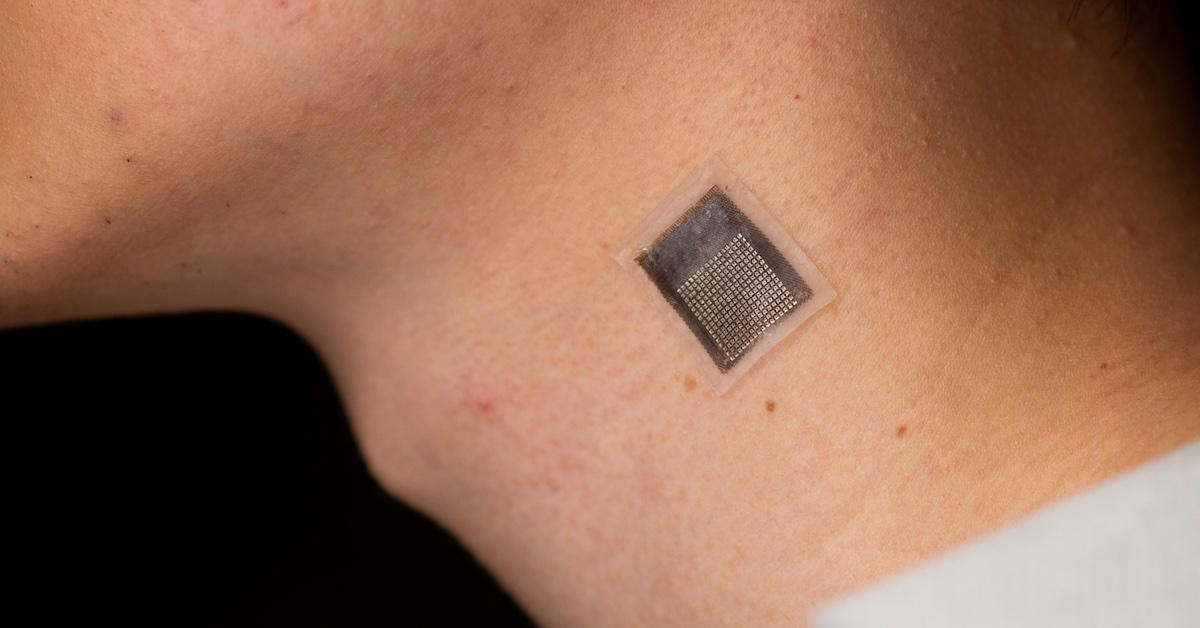
Ultrasound patch worn on the neck. Credit: UC San Diego Jacobs School of Engineering
Improved measurement of tissue stiffness may result in higher therapies for varied circumstances, together with most cancers and sports activities accidents.
A bunch of engineers on the University of California San Diego has created a stretchable ultrasonic array that may carry out non-invasive, serial 3D imaging of tissues as deep as 4 centimeters under the floor of the human pores and skin. This progressive methodology boasts a spatial decision of 0.5 millimeters and presents a extra prolonged, non-invasive answer in comparison with present methods, with enhanced penetration depth.
The examine comes from the laboratory of Sheng Xu, a Professor of Nanoengineering at UC San Diego’s Jacobs School of Engineering and the lead creator of the analysis. The findings had been just lately revealed within the journal Nature Biomedical Engineering.
“We invented a wearable device that can frequently evaluate the stiffness of human tissue,” mentioned Hongjie Hu, a postdoctoral researcher within the Xu group and examine coauthor. “In particular, we integrated an array of ultrasound elements into a soft elastomer matrix and used wavy serpentine stretchable electrodes to connect these elements, enabling the device to conform to human skin for serial assessment of tissue stiffness.”
The elastography monitoring system can present serial, non-invasive, and three-dimensional mapping of mechanical properties for deep tissues. This has a number of key purposes:
- In medical analysis, serial information on pathological tissues can present essential data on the development of ailments comparable to most cancers, which usually causes cells to stiffen.
- Monitoring muscle tissue, tendons, and ligaments will help diagnose and deal with sports activities accidents.
- Current therapies for liver and cardiovascular diseases, together with some chemotherapy brokers, might have an effect on tissue stiffness. Continuous elastography may assist assess the efficacy and supply of those drugs. This may help in creating novel therapies.
In addition to monitoring cancerous tissues, this know-how may also be utilized in different eventualities:
- Monitoring of fibrosis and cirrhosis of the liver. By utilizing this know-how to guage the severity of liver fibrosis, medical professionals can precisely observe the development of the illness and decide probably the most applicable course of therapy.
- Assessing musculoskeletal problems comparable to tendonitis, tennis elbow, and carpal tunnel syndrome. By monitoring modifications in tissue stiffness, this know-how can present useful perception into the development of those circumstances, permitting medical doctors to develop individualized therapy plans for his or her sufferers.
- Diagnosis and monitoring for myocardial ischemia. By monitoring arterial wall elasticity, medical doctors can establish early indicators of the situation and make well timed interventions to forestall additional harm.
Wearable ultrasound patches accomplish the detection operate of conventional ultrasound and likewise break by way of the restrictions of conventional ultrasound know-how, comparable to one-time testing, testing solely inside hospitals and the necessity for employees operation.
“This allows patients to continuously monitor their health status anytime, anywhere,” mentioned Hu.
This may assist scale back misdiagnoses and fatalities, in addition to considerably chopping prices by offering a non-invasive and low-cost different to conventional diagnostic procedures.
“This new wave of wearable ultrasound technology is driving a transformation in the healthcare monitoring field, improving patient outcomes, reducing healthcare costs and promoting the widespread adoption of point-of-care diagnosis,” mentioned Yuxiang Ma, a visiting pupil within the Xu group and examine coauthor. “As this technology continues to develop, it is likely that we will see even more significant advances in the field of medical imaging and healthcare monitoring.”
The array conforms to human pores and skin and acoustically {couples} with it, permitting for correct elastographic imaging validated with magnetic resonance elastography.
In testing, the system was used to map three-dimensional distributions of the Young’s modulus of tissues ex vivo, to detect microstructural harm within the muscle tissue of volunteers previous to the onset of soreness and monitor the dynamic restoration means of muscle accidents throughout physiotherapy.
The system consists of a 16 by 16 array. Each ingredient consists of a 1-3 composite ingredient and a backing layer constructed from a silver-epoxy composite designed to soak up extreme vibration, broadening the bandwidth and bettering axial decision.
Reference: “Stretchable ultrasonic arrays for the three-dimensional mapping of the modulus of deep tissue” by Hongjie Hu, Yuxiang Ma, Xiaoxiang Gao, Dawei Song, Mohan Li, Hao Huang, Xuejun Qian, Ray Wu, Keren Shi, Hong Ding, Muyang Lin, Xiangjun Chen, Wenbo Zhao, Baiyan Qi, Sai Zhou, Ruimin Chen, Yue Gu, Yimu Chen, Yusheng Lei, Chonghe Wang, Chunfeng Wang, Yitian Tong, Haotian Cui, Abdulhameed Abdal, Yangzhi Zhu, Xinyu Tian, Zhaoxin Chen, Chengchangfeng Lu, Xinyi Yang, Jing Mu, Zhiyuan Lou, Mohammad Eghtedari, Qifa Zhou, Assad Oberai and Sheng Xu, 1 May 2023, Nature Biomedical Engineering.
DOI: 10.1038/s41551-023-01038-w
The examine was funded by the Air Force Research Laboratory and the National Institutes of Health.
Professor Xu is now commercializing this know-how through Softsonics LLC.
[adinserter block=”4″]
[ad_2]
Source link